Showing posts with label Dissection. Show all posts

Tuesday, October 6, 2015

Northern Goshawk necropsy; Aspergillosis and Trichomonas

I saw a necropsy of a juvenile wild Goshawk (Accipiter gentilis gentilis) today, which sadly died after being brought into the clinic last week.

He had anklets put on his legs so that he could be perched and fed on the glove but was found collapsed on Sunday afternoon.

The bird died from chronic Aspergillosis seen as spores and large granulomas in the air sacs.
They think the bird had been ill for several weeks as the granulomas were so big and that would coincide with his capture and being brought into the clinic.


He also had secondary Trichomonas gallinae parasites seen in the mouth which were probably elevated due to immunosuppression.

Friday, January 20, 2012

Dissection: Mare Reproductive Tract

Last semester we dissected a Mare's reproductive tract, which was interesting because she was on oestrus.
As with all the dissections, we had to write it up but this time we couldn't just use the photos, we had to draw what we saw in the dissections and put them in the assignment ... was actually quite hard to draw them so had to try + trace it, esp in the proper scientific style! Looking at them now I think they look quite good once they were scanned in, didn't think so at the time!

So we started off with the tract, cut away the fat and bits we didn't need and got:
Dissected the vulva and vagina distally to see the interior + the cervix which is a folded smooth muscle with an interior cervical canal down the middle!

Dissected Mare reproductive tract:


 Cervix:

Then, moving proximally (up!) we found the oviducts and the utero-tubal junction + infundibulum:
Then we get to the ovaries, there were 2, one in oestrus and one in anoestrus.
So first the outside of the ovary:
Then a cross section of an ovary in oestrus (reproductively active mare) - you can see the follicles and corpus luteum:

Tuesday, November 16, 2010

Dissection: Equine Head

Did another equine dissection today ... the head.

We got in and there were heads on the tabled cut off at the top of the head and the ears had already been cut off.
*rank photo warning* ... just incase! ;)
The horses weren't killed for the practical, they are from ill horses which have been put down etc ... people do ask - just saying 'cos people can get stressed/upset by it!
Started off with the head in the tray:

Started off by cutting the skin from the top of the head, right down the nose to get the skin off, slowly cut it off, around the eyes,  turned it upside down and got the skin off in one piece.

Had a look at the muscles, cut a few off to look at them + compare the different ones
... the big one (masseter) is for munching side to side and the smaller one on the right is the temporalis muscles for biting the jaw shut.
Surprised me that the temporalis were so much smaller esp when you think about how hard they can bite!


We had a look at the horses teeth and guessed it was about 2 years old from the angle they meet, how worn down they were and from the lines and stars on the teeth.


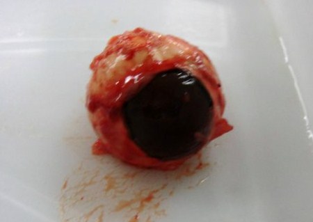
Next had a look at the ear and Becky cut through it to get into the inner ear and I around the eye socket and got the eye out. We saw all the muscles and ligaments around the eye and saw the optic nerve - a little white nerve coming out the back. I cut away everything around it so we just the the eyeball and cut it in hald; was full of a clear liquid with bits of black in it, then there was a lump that was like yellow jelly, and a clear sphere in the middle like one of those bath beads - the lens! 8) If you touched the inside the black/blue lining was soo delicate, literally just fell apart..


We cut the top jaw in half with a hack saw so we could see inside the nasal cavities, have a better look at the teeth, pallet etc.
The pallet was actually soft and a bit rubbery, not like the roof of our mouthes ... can also see the 'stars' in the bottom of the teeth.

Next cut open the top of the skull and got the brain out!!
It was so weird to see what you typically expect from cartoons.
It literally came apart as you held it - pulled the 2 hemispheres apart. It was really soft, as if it was made out of creme cheese!! Soo amazing that people can do brain surgery on something so delicate as that.
Cut one of the hemispheres in half to have a look inside:

I thought it was all really interesting and got really stuck in!

Was the best practical and dissection we've done, although some people were a bit squeamish about it - I could have cut away and dissected it all for hours though! :P

Friday, November 5, 2010

Dissection: Reproductive tract of a cow

We had another lab practical today ...
Dissection of the reproductive tract of a cow.

*rank photo warning* ;)

There were others in there that we had a look at too; mare, ewe lamb and sow.

We had a bit of digestive tract attached to ours so it stank.
What we got in the tray at the start:


I was shocked at how much fat there was around it too!
Probably helps insulate it, but half of it was fat that we had to cut away at the start.

Can see top right all the fat that we cut away!


We found everything we were looking for, then started at the vagina, dissected all the way down the cervix + to the ovaries and there was a follicle at the end. The follicle was a little sac a couple of cm big and inside it was the ovary which is 1/10th of a mm!
Not a great photo, but the follice:

Cows ovaries:

All labelled up at the end: